Posted on February 15, 2021
Two University of Pretoria (UP) veterinary surgeons have performed life-saving heart surgery on two dogs by using a ground-breaking technique. This is a first in the 100-year history of the Faculty of Veterinary Science in Onderstepoort, and an exciting way to start the next century of veterinary service to the country.
Dr Adriaan Kitshoff and Dr Ross Elliott, who are specialist veterinary small animal surgeons working in Small Animal Surgery in the Department of Companion Animal Clinical Studies, saved the lives of 7-month-old French Bulldog Daisy and a 6-month-old Cocker Spaniel called Tallen. Daisy is a service dog that can sense when her owner, who has fibromyalgia, is in pain and sleeps on her as a means of comfort.
The vets used a ground-breaking approach that entails dilating the opening of a heart valve with a balloon. “This procedure is limited to hospitals overseas with surgeons or internists with special interest in cardiology. It is only hospitals that have the equipment that can perform the surgery,” they said.
They explained that both their patients have pulmonic valvular stenosis, an abnormally shaped or fused heart valve that is situated between the heart and the artery leading to the lungs. “Both these patients had valves where a component had fused together. These valves are supposed to allow the blood to flow in one direction, meaning that they have times that they are closed and times that they are open and these times are determined by whether the heart contracts or not.”

Post-operative Daisy during her stay in the intensive care unit of the Onderstepoort Veterinary Academic Hospital.
In the case of valves that are fused, the valve can’t open properly when the blood should leave the heart (during contraction) and also does not close properly (when the heart relaxes). “This created a scenario that is similar to you filling your lungs with air and blowing out through your mouth with your lips pursed. The force of the heart trying to squeeze out blood through a small opening places tremendous strain on the heart muscle.”
The aim of the surgery for both dogs was to increase the size of the opening by dilating the valve. For this, a long balloon-tipped catheter was placed in one of the neck veins. Through fluoroscopy (“real-time X-rays”), the balloon was guided through two of the heart chambers and through the small opening in the valve. After inflation of the balloon the opening was stretched (balloon valvuloplasty).
The vets explained that the anatomy of a dog’s heart is similar to that of a human’s, also consisting of four chambers and four valves. “This is probably the reason why most of the clinical trials for heart transplants were performed on dogs. This set the stage for Dr Chris Barnard’s first successful heart transplant in 1967.”
The surgery is risky as these patients have heart disease and need to be placed under general anesthesia. Due to irritation of the heart muscle as the balloon passes through it, it can result in abnormal heart rhythms during the anesthesia that might need to be treated with medication during the operation. Stretching of the valve can cause tearing of some blood vessels.

LEFT: Dr Ross Elliott and Sister Adele Rossouw doing a dissection out the neck vein (jugular vein) for insertion of the balloon catheter. RIGHT: Dr Adriaan Kitshoff performing fluoroscopy to assess the location of a guide wire used during the balloon catheter placement.
Furthermore: “These abnormal rhythms and tearing of blood vessels can be fatal in a very small percentage of patients. In some dogs the opening between the fused valve can be so small that it is not possible to pass the deflated balloon through it. In these patients the chest needs to be opened up, a hole needs to be made in the beating heart and an instrument passed into the heart to stretch the opening of the valve.”
The implications of the procedure performed are that “we can offer a service not previously offered by Onderstepoort Veterinary Academic Hospital. This might be the start of other minimally invasive heart surgeries in the future. This also provides us with the opportunity to extend the life of special pets like these.”
This ground-breaking surgery also creates the possibility of setting up a centre of excellence in minimal invasive surgery and cardiology at the hospital and offering more advanced surgeries like valve replacements and heart transplants.
Both dogs are doing well after their surgeries but need follow-up heart scans every three months as in 15% of such cases the stretched opening of the valve can start to narrow again. “In which case the procedure can be repeated again,” the vets said. The surgery was a team effort which included anesthetists Dr Justin Grace and Dr Abdur Kadwa, theatre nurse Sister Adele Rossouw and theatre assistant Mike Shabangu.
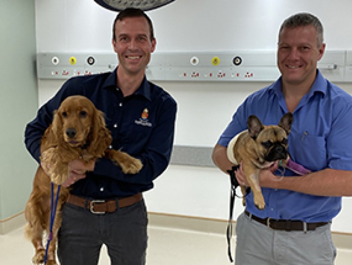
Specialist surgeons Dr Adriaan Kitshoff and Dr Ross Elliott with Tallen and Daisy.


Copyright © University of Pretoria 2025. All rights reserved.


 Virtual Campus
Get Social With Us
Download the UP Mobile App