


Posted on July 26, 2021
The Onderstepoort Veterinary Academic Hospital (OVAH) of the University of Pretoria’s (UP) Faculty of Veterinary Science has acquired its first magnetic resonance imaging (MRI) scanner. This means that the hospital now no longer has to make special arrangements with human hospitals to perform MRI scans.
“In South Africa, the use of MRIs for diagnostic purposes is limited to small animals, and most MRI scans are done in human facilities by special arrangement,” said Dr Paul van Dam, Director of the OVAH. “An MRI scan takes 45 minutes or longer, which limits the number of cases that can be referred. With our own MRI scanner, we can now do scans at any time of the day, on site, without the additional time to travel to another facility. An added advantage is that our MRI scanner will be the only high-field MRI in South Africa, and probably Africa, with image acquisition optimised for veterinary patients.”
In the past, the OVAH has facilitated MRI scans on a variety of species, including several highly trained working dogs and a lion.

A dog being scanned on the new high-field MRI scanner.
MRI scanners differ in strength in terms of the magnetic field being used, which is rated in tesla (T) units. Stronger magnetic fields make for faster examinations, with greater detail and clearer pictures compared to lower-strength units. “The new MRI scanner at the OVAH is a 1,5T unit, which is believed to be the strongest unit in veterinary use anywhere in Africa,” Dr Van Dam said. “The unit also has the biggest diameter bore (the scanner’s diameter) available, which allows us to scan the bodies of even the largest dogs and many other animals – including wildlife such as big cats and great apes – on the built-in table. We can also scan the limbs and necks of larger animals like horses on a purpose-built (and locally designed) table.”
He explained how an MRI works. “Magnetic resonance imaging is the newest form of imaging in general use today. Through this method, the molecules in the body resonate when a magnetic field (of up to 60 000 times as strong as the magnetic field of the earth) is applied to them. A short pulse of radio waves is applied, and a weak signal is echoed back from the body tissues; the echo varies depending on the type of tissue involved.”
MRI scanners have been used in human medicine since 1977 and have been used in veterinary medicine since the mid-1990s. MRI imaging is the diagnostic method of choice when there is low contrast between neighbouring tissue types. This includes the brain – where the MRI distinguishes between grey matter, white matter, nerves and cerebrospinal fluid – as well as the locomotor system, where it can distinguish between muscle, tendons and ligaments surrounding joints, joint fluid and joint cartilage. Lesions of the spinal cord are also best visualised with MRI imaging.

Student Lulama Nsele with Bear, a Yorkshire terrier, the first patient in the MRI facility.
“By providing excellent images of soft tissue, MRI scans enable the clinician to make an accurate and often early diagnosis, allowing properly targeted treatment plans,” said Dr Van Dam.
The OVAH has the largest variety of veterinary specialists practicing under one roof out of all veterinary hospitals in South Africa. These veterinarians examine, diagnose and treat patients supported by veterinary sisters who provide specialised nursing care. Enthusiastic students assist the clinical team as part of their practical training.
The hospital boasts an array of facilities and equipment, including an extended and improved state-of-the-art dog and cat ICU and high-care facility, 10 theatres for small and large animals where total hip replacements, implant-driven orthopaedic procedures for cats and dogs and life-saving emergency surgery on all animal species are performed. Internal medicine, including oncology, neurology and dermatology is practiced in species-specific clinics and the equine clinic is equipped to diagnose and treat complicated lameness cases.
Facilities also include accommodation for all animal species, from the smallest pet to horses, farm animals and wildlife, including the big cats and even immature rhinos. Added to this, the OVAH has access to a number of specialised on-campus diagnostic laboratories and specialist pathology services, enabling it to give each patient the best individual care, attention and treatment. “All this ensures that the OVAH is never lacking in patients to heal or opportunities to train students to the highest standards,” said Dr Van Dam.

Wiskey the dog is being prepared for a scan with Sister Sinazo Nikelo assisting.
“The acquisition of the MRI scanner provides the hospital with a comprehensive set of imaging modalities including conventional radiographs (X-rays), ultrasound, scintigraphy, computed tomography (CT) and now magnetic resonance imaging (MRI),” said Professor Vinny Naidoo, Dean of the Faculty of Veterinary Science at UP. He explained that the MRI scanner was made possible in part by a generous bequest from Betty Noakes, who left a large part of her estate to the OVAH to be used for the benefit of animals. “We trust that the MRI will indeed play a large role in improving the life of our patients.”
Professor Tawana Kupe, Vice-Chancellor and Principal of UP, who officially opened the new facility, also acknowledged the importance of the new acquisition. “Over the years, the OVAH has distinguished itself on various levels. It has now begun another exciting chapter in the faculty’s more than 100-year history – the beginning of a new era in service delivery and patient care. It reinforces the hospital’s invaluable role and position as the country’s leading veterinary hospital.”
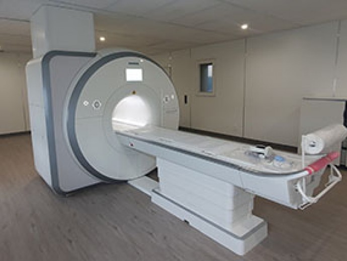
The high-field MRI scanner of the Onderstepoort Veterinary Academic Hospital, believed to be the first of its kind in Africa.

Copyright © University of Pretoria 2025. All rights reserved.
 Virtual Campus
Get Social With Us
Download the UP Mobile App