What do you do when a vulture with a crushed beak needs a new beak and two attempts to fit an acrylic beak fail? You improvise and use the beak of a deceased vulture, successfully enabling the injured bird to feed again.
In another first for South Africa, this remarkable procedure was recently performed on a white-backed vulture (Gyps africanus) by a University of Pretoria (UP) team led by Professor Katja Koeppel, a veterinary wildlife specialist in UP’s Faculty of Veterinary Science at the Onderstepoort campus.
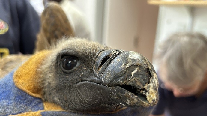
The beak of the female African white-backed vulture, an endangered species, was crushed when it was hit by a car in March this year. The bird also presented with a serious head injury, causing blindness in the right eye. The vulture was taken in by non-profit conservation organisation VulPro, and was hand-fed as it was not able to feed independently at the time.
VulPro CEO Kerri Wolter then brought the bird to UP’s Faculty of Veterinary Science, where Prof Koeppel, together with wildlife veterinarians Dr Jennie Hewlett and Dr Bart Gazendam, veterinary nurse, Sr Murendeni Lalamani and a few students, attempted on two occasions to fit the bird with prosthetic acrylic beaks and wires. However, the vets found that the prosthetic became unstable once the vulture tried to feed on a carcass.
Following a more innovative approach, Prof Koeppel then decided to use an intact beak from a deceased vulture; this would provide the correct shape for tearing meat. Fortunately, a matching piece of beak was obtained from a dead white-backed vulture which was stored in a freezer for research purposes. Prof Koeppel and the team were able to perform the ground-breaking procedure in May, which involved using six orthopaedic screws to attach the beak, thus saving the bird’s life.
The result has been remarkable.
“The vulture has shown tremendous improvement,” Wolter said. “She is doing fine and is eating well with the new beak. The crushed beak, which is made of keratin, will continue to grow back fully underneath the bit bolted onto it, but it will probably take a few years. The transplanted beak will then be taken off.”
The vulture will be staying at VulPro and be monitored on a continuous basis. Unfortunately, the blindness caused by the head injury means that she will not be able to rely on her otherwise excellent sight any longer, and will not be released back into the wild.
** The white-backed vulture is the most common and widespread vulture species in sub-Saharan Africa but is listed as an endangered species because of a decreasing population. These scavengers are vital to our ecosystem as they feed primarily on the carcasses of dead animals, thus clearing the landscape of carrion and helping to curb the spread of dangerous diseases and bacteria.
Click on the gallery in the sidebar to view pictures on the surgery process.
Professor Katja Koeppel
June 9, 2023
 Story
Story
Cricket à la king? How about a yellow mealworm burger? Foods that may previously have evoked a ‘yuck’ response are now firmly on the menu. Research into edible insects by the Department of Zoology and Entomology at the University of Pretoria (UP) is exploring how to rear and harvest this food of the future.
 Story
Story
Generative artificial intelligence (GenAI) is changing how we work, play and relax. Whether you use ChatGPT to write a brief, Midjourney to generate visuals or MuseNet to create unique soundtracks, these technologies have opened up opportunities for richer content.
 Story
Story
A single query to ChatGPT uses as much electricity as burning a light bulb for about 20 minutes. Multiply that by the millions of requests that this artificial intelligence (AI) chatbot receives each day, and the environmental impact is ominous.
Copyright © University of Pretoria 2025. All rights reserved.

Get Social With Us
Download the UP Mobile App