
Posted on February 13, 2024
We are thrilled to share some big (pun intended) news from the Malaria Parasite Molecular Laboratory (M2PL), as part of the University of Pretoria Institute for Sustainable Malaria Control (UP ISMC). We successfully accomplished the first-ever ultra-structure expansion microscopy (U-ExM) of malaria parasites in South Africa, a milestone for the malaria research community in Africa.
Malaria, a global health concern, disproportionately affects populations in Africa, and innovative approaches are crucial to unravelling the biological complexities of the parasite that causes this disease. The Plasmodium parasite is so small that it fits into one of the human’s smallest cells, red blood cells, which is ~8 μm in diameter. Indeed, during its development, the parasite is as small as ~1.5 µm and maximally reaches ~5 µm. This poses challenges for using conventional fluorescent, confocal microscopy to study the parasite morphologically. Even with the amazing breakthroughs provided by super-resolution microscopy, much of the parasite’s biological structures are still difficult to observe or examine.
U-ExM is a pioneering microscopy technique that aims to expand biological specimens physically, making them larger and easier to visualise. This entire idea was a translational invention from the Massachusetts Institute of Technology (MIT) in the US based on the expansion associated with baby diapers when wet! U-ExM can enlarge (expand) a biological structure ~10 fold linearly while retaining the 3D context and intracellular connectivity without requiring any additional resolution power from the microscope itself. This lends itself perfectly to studying small, single-celled organisms such as the malaria parasite (Figure 1).

Figure 1: How we normally see the smallest version of the immature malaria parasites (merozoite) using conventional light microscopy (right). U-ExM has the ability to show us interacted details within a single merozoite (left).
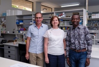
The application of U-ExM to malaria parasites allowed us to achieve an unparalleled level of resolution for fluorescent samples instead of using electron microscopy. Henrico Langeveld, a PhD student under the supervision of Prof Lyn-Marié Birkholtz as director of the M2PL, in collaboration with the Africa Microscopy Initiative (AMI), situated at the Institute for Infectious Disease and Molecular Medicine (IDM) at UCT, applied U-ExM to study different stages of malaria parasites.
The U-ExM enlarged the parasite ~5 fold, allowing us to visualise intracellular structures with unprecedented clarity and detail (Figure 1). Using various structure-specific fluorescent dyes and antibodies, we could showcase intricate details of the parasite's cellular structures during different parasite stages, something not observed before (Figure 2). To our knowledge, this is the first-ever experiment of this nature performed in Africa.

Figure 2: Top panel: conventional light microscopy and fluorescently labelled mature malaria parasites. Super resolution (SR) fluorescent microscopy showcases some of the parasite’s insides (green = nuclei, red = tubulin). Bottom panel: After U-ExM the detail is staggering to the point where we can see individual tubulin stands (cyan = nuclei, magenta = tubulin). U-ExM allows us to visualise the microtubule organising centre (MTOC), which is not visible when using conventional light microscopy or SR fluorescent microscopy.
Performing and establishing this technique in Africa holds significant local relevance. Our work not only contributes to the global scientific community but also addresses the specific challenges and characteristics of malaria strains prevalent in African regions. This technique will answer current outstanding questions and open up new avenues for research and potential breakthroughs in treatment strategies and unravelling the unique biology of the parasite.
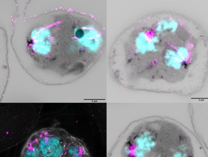
Images of malaria parasites taken after ultra-structure expansion microscopy (U-ExM)

Copyright © University of Pretoria 2024. All rights reserved.
 Virtual Campus
Get Social With Us
Download the UP Mobile App