
Posted on April 26, 2024
In the realm of medicine and research, technological advancements continue to revolutionize the way we understand and treat conditions.
CT scans have been around for more than 50 years and are used to show detailed images of various parts of the body, including bones and organs. CT scans are made up of many cross-sectional images, which are usually looked at individually in the medical field. More recently, the ability to transform CT scans into physical 3D models has allowed much better visualization of the scan and offers profound insights into the anatomical intricacies of the patient. CT scans are also often used in the veterinary field. The use of a 3D model improves treatment planning and serves as a guide for surgical interventions.

3D Model with Internal Detail
The Library MakerSpace was involved in a project to create a 3D model based on a CT scan done on a dog’s heart. This model was created for the commissioning of the new CT scanner that was recently acquired by the Veterinary Hospital.

3D Model Print Preview
This project exemplifies the MakerSpace's capacity to support university research with remarkable results.
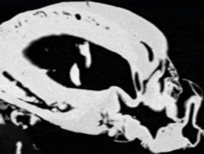
The MakerSpace technicians processed the raw data from the CT scan, incorporating all internal details to produce a usable 3D model.


Copyright © University of Pretoria 2025. All rights reserved.
 Virtual Campus
Get Social With Us
Download the UP Mobile App